Medanets and imito help elevate wound documentation

Traditional wound documentation often relies on manual measurements and using multiple devices, such as cameras and computers. This is time-consuming and prone to human error. What if you could accelerate the process and improve accuracy at the same time?
Medanets, a health company from Oulu, partners with the Swiss imito Ltd. to enhance the wound documentation process with artificial intelligence.
The traditional process of wound documentation
Imagine you had a patient with a wound, and every detail of their wound needed to be documented manually. You start by documenting the type and origin of the wound, its exact location, and how long it has been there. You then grab a disposable ruler to measure the wound’s length and width, carefully jotting down the dimensions. Or, if you don’t have a ruler available, you start to think of commonly known shapes and sizes, such as ‘the size of a one penny coin’. If there’s a cavity, you measure and document the direction, perhaps noting a cavity at 3 o’clock.
As you observe the wound bed, you note the types of tissue present — red granulation tissue, perhaps. You also record the amount and type of drainage and any redness or swelling around the wound. Then comes the pain assessment, asking the patient to rate their discomfort and noting if it worsens during dressing changes.
Finally, you apply the appropriate dressings, carefully documenting every step. This process repeats regularly as you track the healing progress. Perhaps you need to change the expensive dressings to show the recently treated wound to a doctor. All the while, the process consumes valuable time and other resources.
Did you have a laptop in the room where you assessed the patient’s wound and skin, or did you write all this down on paper just to type it again into the Electronic Health Record? Maybe you had a camera with you, took a picture of the wound, then transferred it into the computer, and remembered to delete the picture from the camera’s local memory?
The streamlined process with Medanets & imito
Now, imagine the same patient again. You open the Medanets app on your mobile device, start the documentation process by identifying the patient electronically, and open the Clinical photos feature. The app gives you structured alternatives when documenting the type, location, and other vital data regarding the wound.
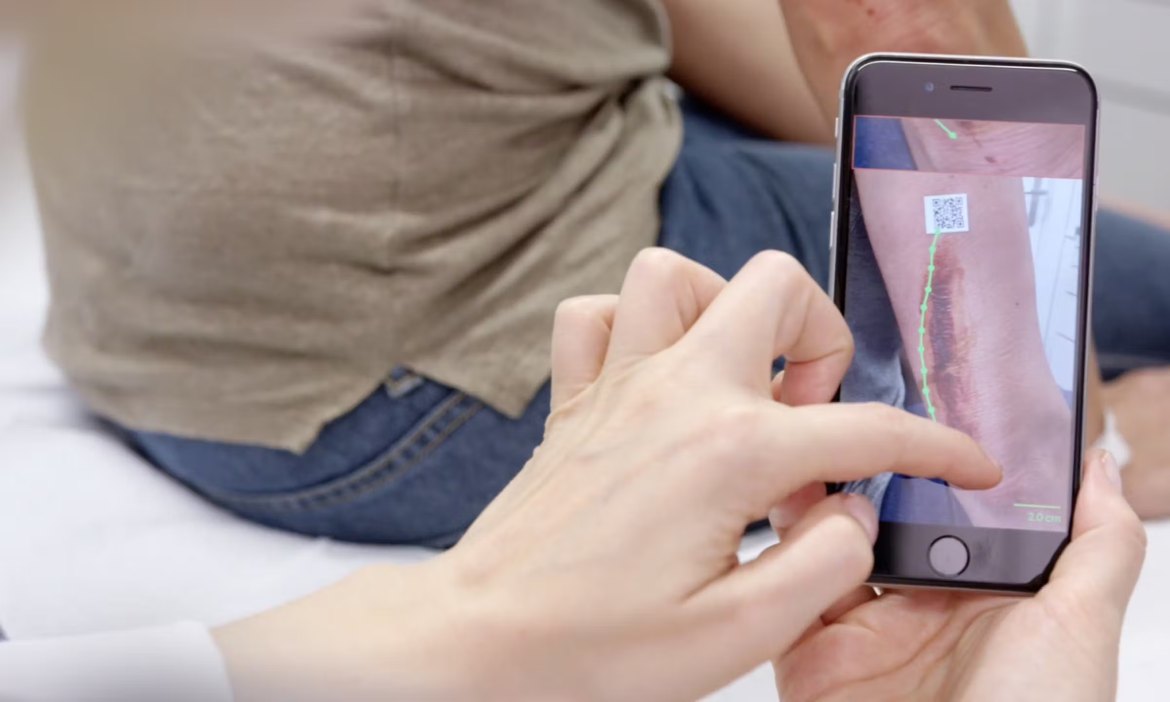
Then, you place a calibration marker next to the wound and proceed to take a picture, still using the same mobile device. You are seamlessly transferring from Medanets to the imitoWound solution without separate logins or other nuisances. The solution automatically considers the lighting conditions and adjusts the camera to suit them. You can skip manually measuring the wound altogether, as the solution measures the wound precisely and automatically. When you take a picture, Imito automatically detects the wound’s edges and calculates the surface area. You can also monitor the healing of the wound by comparing the new and old images and measurements directly in the app.
imito allows multiple images to be taken, stored, and transferred to the image archive simultaneously. You can also view the images in imitoWound and decide which ones to send to the image archive.
Wound documentation becomes highly automated and fully standardised. Once stored, the images will be immediately available to everyone involved in the treatment.
Soon, imitoWound will be further enhanced with AI tools, such as tissue segmentation and 3D reconstruction of the wound, for improved diagnostic accuracy. All this optimises patient outcomes and reduces risks through informed treatment decisions.
Medanets is currently looking for pilot customers. If you are interested, do not hesitate to contact sales@medanets.com!
Source: Medanets
Image: Imito
